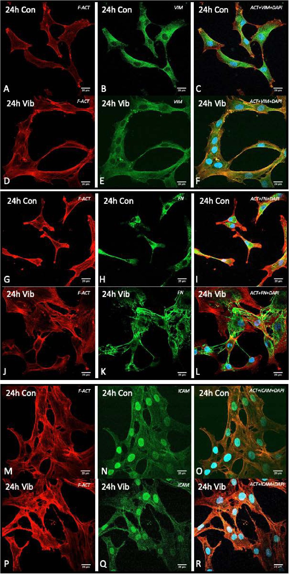
Fig. 2. Immunofluorescence staining of vimentin (B, E), fibronectin (H, K) and ICAM-1 (N, Q) (green), F-actin filaments (red; A, D, G, J, M, P) and the nucleus (blue - DAPI) in chondrocytes exposed to static control conditions (A-C, G-I, M-O) or to VIB (D-F, J-L,P-R) for 24 h. The scale bar is 20 µm.